The Nervous System
Science TEAS Review – The Nervous System
The previous lessons in the science TEAS review covered the organization of the body systems. Now we discuss integration and control with this science TEAS lesson introducing the anatomy of the nervous system, including its functions and divisions. It also explores the parts of neuron, neural conduction, and synaptic transmission.
The following videos will provide a review on the Nervous System.
Part I: Division of the Nervous System
Part II: Neurons and the Synapse
Science TEAS Review – What Is the Nervous System?
From perceptions to daily experiences, the nervous system controls many aspects of the human body. This system coordinates several activities in the body. It governs people’s consciousness, their personalities, how they learn, and their ability to memorize. Working with the endocrine system, the nervous system regulates and maintains homeostasis.
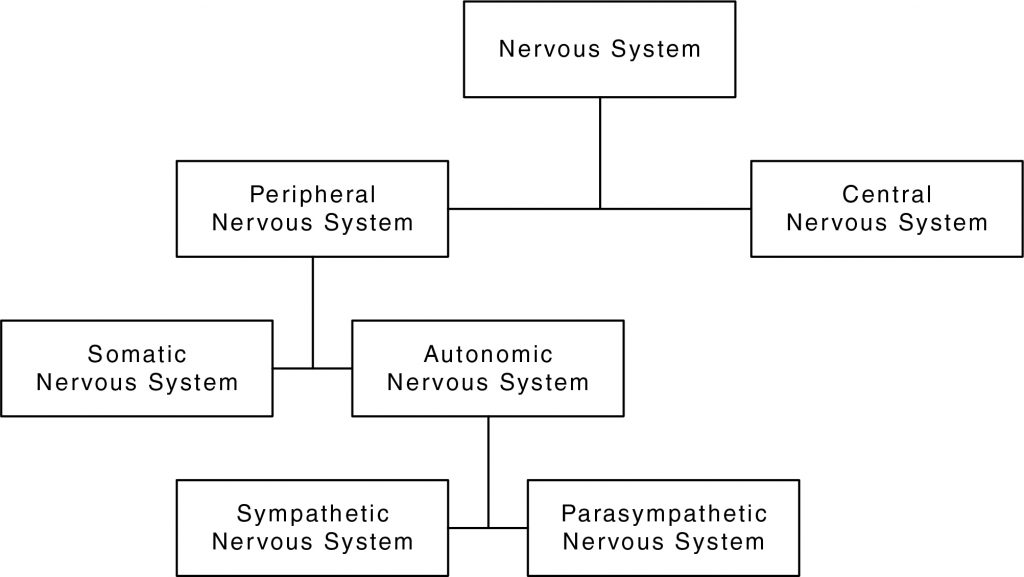
The following flow chart summarizes the divisions of the nervous system.

The nervous system is anatomically divided into two parts:
- Central nervous system (CNS): The central nervous system is comprised of the brain and spinal cord. It is where information processing and control occurs.
- Peripheral nervous system (PNS): The peripheral nervous system is comprised of the nerves associated with the CNS. It connects all nerves of the body to the CNS. There are two types of fibers in the PNS: (a) afferent fibers that transmit impulses from organs and tissues of the body to the CNS; and (b) efferent fibers that transmit impulses from the CNS to the organs and tissues of the body.
The PNS is further divided into the somatic and autonomic nervous systems. The somatic nervous system primarily controls voluntary activities such as walking and riding a bicycle. Thus, this system sends information to the CNS and motor nerve fibers that are attached to skeletal muscle. The autonomic nervous system is responsible for activities that are non-voluntary and under unconscious control. Because this system controls glands and the smooth muscles of internal organs, it governs activities ranging from heart rate to breathing and digestion. The autonomic nervous system is further divided into the following:
- Sympathetic nervous system: The sympathetic nervous system focuses on emergency situations by preparing the body for fight or flight.
- Parasympathetic nervous system: The parasympathetic nervous system controls involuntarily processes unrelated to emergencies. This system deals with “rest or digest” activities.
Based on the activities of the nervous system, this system can be functionally divided into three parts:
Test Tip

The first letter in the parasympathetic and sympathetic nervous system can be used to tell them apart:
Sympathetic = stress
Parasympathetic = peace
- Sensory: Information is gathered (both internally and externally) and carried to the CNS. The senses gather the information that the sensory nervous system transmits.
- Integrative: The integrative nervous system is where the CNS process and interprets information received from the sensory nerves.
- Motor: Motor nerves convey information that is processed by the CNS to muscles and glands.
Try this practice TEAS science test question
Science TEAS Review – Anatomy of the Brain
The brain is a mass of tissue that is made of billions of nerve cells called neurons. This complex organ controls a wide range of processes and integrates information received from the five senses. Protected by the skull, the brain consists of four cavities called ventricles. These cavities are filled with cerebrospinal fluid (CSF), which surrounds the CNS. This fluid serves many purposes such as protecting the brain from physical shocks and removing wastes from the neural tissue in the brain.
Keep In Mind

The lobes are named after the bones of the skull that protect each lobe. For example, the frontal bone protects the frontal lobe.

As shown in the above image, the brain is divided into the following three regions:
- Cerebellum: This is found beneath the cerebrum and behind the brainstem. It helps coordinate body movements, posture, and balance.
- Brainstem: This is found between the thalamus and the spinal cord. It is the lowest part of the brain that connects the brain with the spinal cord. Unconscious functions like breathing, heart rate, and blood pressure are controlled by the brainstem.
- Cerebrum: This part of the brain is the largest and part of the forebrain. The cerebrum controls higher-order functions such as interpreting touch, speech and language, reasoning, emotions, and fine motor control.
The cerebral cortex is grey (or gray) matter that surrounds the entire cerebrum. It is divided into a left and right hemisphere. The ridges of the cerebral cortex are called gyri, and the grooves are called sulci. The very large grooves are called fissures. The cerebral cortex is divided into four lobes: the frontal, parietal, temporal, and occipital lobe.
The cerebral cortex is the most complex part of the brain, and each lobe has specific functions that are outlined in the following table.
| Lobe | Function |
| Frontal | Processes high-level cognitive skills, reasoning, concentration, motor skills, language, and functions as a control center for emotions. |
| Parietal | Integration site for visual perception and sensory information such as touch, pain, and pressure. |
| Temporal | Organizes sounds and processes language that is heard. Helps form memories, speech perception, and language skills. |
| Occipital | Interprets visual stimuli and information. |
Try this practice TEAS science test question
Science TEAS Review – The Thalamus and Limbic System
Recall that the cerebral cortex is composed of grey matter. This mater is a type of neural tissue that contains three types of neurons, which are nerve cells that make up the nervous system:
- Sensory neurons: Afferent nerve cells that send information toward the CNS. This information is what is sensed, using the five senses, from the external environment.
- Motor neurons: Efferent nerve cells that carry impulses away from the CNS to the effectors, which are typically tissues and muscles of the body.
- Interneurons: Nerve cells that act as a bridge between motor and sensory neurons in the CNS. These neurons help form neural circuits, which helps neurons communicate with each other.

Be Careful!
Grey matter is different from white matter. White matter is found in the spinal cord and surrounds the grey matter. It contains bundles of interneurons.
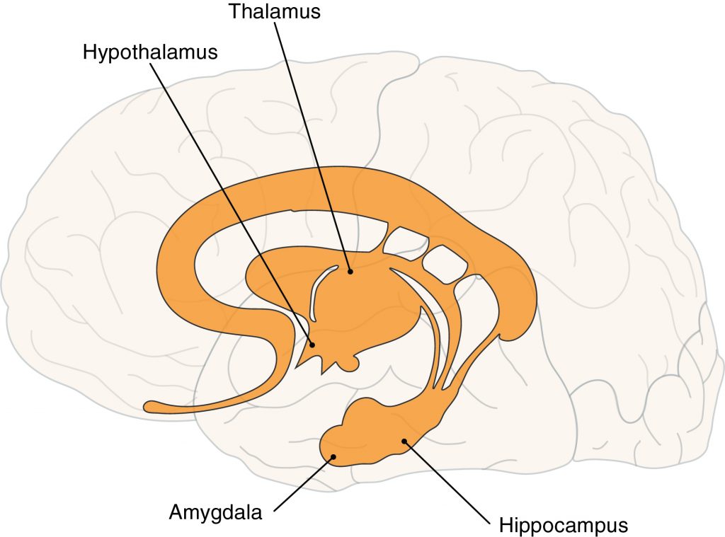
Another part of the forebrain incudes the limbic system, which controls emotions and memory. As shown in the image, this system is found right beneath the cerebral cortex and sits above the brainstem.

Four major structures of the brain comprise the limbic system:
- Hypothalamus: Found below the thalamus, this structure plays a role in regulating the autonomic nervous system. It is primarily concerned with homeostasis and regulates various activities such as hunger, anger, and the response to pain. The hypothalamus works with the pituitary gland from the endocrine system. This gland uses hormones, or chemical messengers, to generate responses in the body.
- Amygdala: Recognized as the aggression center, areas of this region produces feelings such as anger, violence, fear, and anxiety.
- Thalamus: Different sensory inputs come through the nerves and end at the thalamus, which directs this information to various parts of the cerebral cortex. The sense of smell is the only sense that bypasses the thalamus. Information related to movement is also processed by the thalamus.
- Hippocampus: Helps convert short-term memory to long-term memory. If the hippocampus is destroyed, new memories cannot be formed but old memories are retained.
Did You Know?

Kluver-Bucy syndrome is a condition that includes destruction of the amygdala. This means a person will present with erratic emotional behavior symptoms like hypersexuality, compulsive eating, and putting objects in the mouth.
Try this practice TEAS science test question
Science TEAS Review – Anatomy of a Neuron
Recall that the nervous system is comprised of specialized cells called neurons. A large network of neurons work together to quickly send and receive messages throughout the body. As shown in the following image, a neuron has several parts.

A neuron’s structure is designed to transmit electric signals before they are transmitted as chemical signals to a target cell. The following three basic parts make up a single nerve cell:
- Cell body: This is the main part of the neuron that contains the nucleus of the nerve cell. Also called the soma, other organelles are also found in the cell body.
- Dendrites: These are appendages attached to the cell body that receive signals from other neurons.
- Axon: This is the long structure attached to the cell body. It conducts and transmits information to other cells. Branches at the end of the axon form axon terminals. These branches facilitate communication between neurons and target cells.
Be Careful!

Do not confuse a neuron with a neuroglial cell. Neuroglial cells do not conduct nerve impulses like neurons. Rather, they provide support and protect neurons. Astrocytes, oligodendrocytes, microglial cell, and ependymal cells are the four major types of neuroglial cells in the CNS. Schwann and satellite cells are in the PNS.
Also shown in the image is a myelin sheath and node of Ranvier. The myelin sheath is a protein and lipid structure produced by a type of glial cell called a Schwann cell. This sheath functions like a blanket that provides a layer of insulation around the axon of a neuron, increasing the speed of electrical signal transmission. Regularly spaced gaps called nodes of Ranvier are found between the myleinated sheaths. Electric signals jump from one node to the next, thereby increasing the speed of signal transmission.
Did You Know?

Several diseases cause degeneration of the myelin sheath, or demyelination. One example is multiple sclerosis. When demyelination occurs, it can lead to severe neurological problems like motor and cognitive function. Demyelination reduces the speed at which neural impulses are transmitted along the axon.

Try this practice TEAS science test question
Science TEAS Review – Synaptic Transmission and Nerve Impulses
The electric signals neurons transmit are called neural impulses. Neurons must be excited to create a nerve impulse. A stimulus triggers excitation. At the resting state, the inside of the neuron is more negatively charged, while the outside of the neuron is more positively charged. This difference in electrical charge because of potassium and sodium ions establishes the resting potential.

Did You Know?
As a person ages, the rate of neuroplasticity, or ability for the brain to form neural connections through synapses, decreases. Neuroplasticity is important because it helps the brain adapt to new stimulation, damage, or changes in the environment.
During the action potential, a reverse in electrical charge occurs across the membrane of a neuron in its resting state. As shown in the following image, this happens when a neuron receives a neural impulse by way of a stimulus or a chemical signal from another neuron. The inside of the neuron becomes more positively charged, while the outside of the neuron becomes more negatively charged. This reverse in charge travels down the axon as an electric current.

The steps of an electrical synapse are outlined below:
- Once the action potential reaches the terminal bulbs of the axon terminal, the synaptic transmission process begins. The sequential numbers in the image outline the steps of synaptic transmission. The details of each numbered step are outlined below:An action potential travels down the axon and reaches the terminal branches of the axon. Voltage gated sodium gates open, causing sodium to enter the axon terminal bulb.
- Voltage gated Ca2+ channels open at the same time.
- Calcium ions move into the axon terminal bulb of the presynaptic neuron.
- Calcium ions bind with proteins on synaptic vesicles that carry chemical messages called neurotransmitters.
- This binding causes the vesicles to contract and move to the presynaptic membrane.
- Neurotransmitters are released from the vesicles via exocytosis and diffuse across the synaptic cleft.
- Neurotransmitters bind with receptors on the postsynaptic membrane of a neuron, gland, or muscle.
- Depending on what the postsynaptic target cell is, the following responses will happen:
- Axon to dendrite: Action potential travels to next neuron.
- Axon and muscle cell: Muscle contraction.
- Axon and gland: Hormones released from gland.
Try this practice TEAS science test question
Let’s Review
- The nervous system is divided into the central nervous system (CNS) and peripheral nervous system (PNS).
- The somatic nervous system controls voluntary activities, while the autonomic nervous system is responsible for involuntary activities under unconscious control.
- The nervous system performs sensory, integrative, and motor functions.
- The three major regions of the brain are the cerebrum, brainstem, and cerebellum.
- Four lobes comprise the cerebral cortex, which is grey matter that surrounds the cerebrum.
- The limbic system consists of the hypothalamus, thalamus, hippocampus, and amygdala, each of which has different purposes.
- Neurons are made of dendrites, a cell body, an axon, and an axon terminal.
- Myelin sheaths insulate the axon of neuron, increasing the spread of electric signal transmission.
- A neuron must be excited from a stimulus to create a nerve impulse.
- Resting potentials are established when the outside of a nerve cell is more positively charged than the inside of a nerve cell.
- Action potentials are established when the reverse of a resting potential occurs.
- Synaptic transmission occurs in several steps and only occurs following an action potential.
- Neurotransmitters are chemical messengers released during an electrically stimulated synaptic transmission process.
In the next lesson we cover the science TEAS topic for the endocrine system.
Nervous System Flashcards
You May Subscribe to the online course to gain access to the full lesson content.
If your not ready for a subscription yet, be sure to check out our free practice tests and sample lesson at this link
