The Cardiovascular System
ATI TEAS Science – The Cardiovascular System
As you prepare for the ATI TEAS science section, A&P will comprise of 18 questions. This lesson introduces the anatomy of blood and its connection to the cardiovascular system. Reviewing this lesson will allow you to understand the parts that make up the cardiovascular system and how this system functions.
What You’ll Learn and Why It’s Important For Your TEAS 7 Exam
- Identify the components of blood and their functions.
- Explain the process of hemostasis and its significance in wound healing.
- Understand blood grouping and agglutination.
- Describe the anatomy of the cardiovascular system, including the structure of the heart and blood vessels.
- Outline the processes of systemic and pulmonary circulation.
- Interpret an electrocardiogram (EKG) and understand its components.
Need help? Don’t forget Nurse Melissa and more students preparing for the TEAS are just one post away in our TEAS 7 Study Group
Flashcards & Note Prompts
- Create a flashcard for each of the keywords below.
- Create a flashcard or take notes on the elements of blood and their functions. For example, red blood cells participate in gas exchange.
- Create a flashcard or take notes on each of the functions of blood.
- Create a flashcard for the 3 steps of hemostasis
- Create a flashcard for each blood group, specifically whom it can accept blood from and donate blood to.
- Take detailed notes and create a flashcard for each of the heart’s four chambers and the layers of the heart wall.
- Take notes and create flashcards to remember the four valves and their function in blood flow in and out of the heart.
- Take notes or make flashcards on the 4 steps of systemic circulation.
- Take notes or make flashcards on the 5 steps of pulmonary circulation.
- Make flashcards for each wave on an EKG such as the P and T waves.
Keywords and Concepts
- Blood
- Plasma
- Albumin
- Erythrocytes
- Leukocytes
- Thrombocytes
- Viscosity
- Red blood cells
- White blood cells
- Platelets
- Hemostasis
- Blood groups
- Buffy coat
- Agglutination
- Antigens
- Vasodilate
- Vasoconstrict
- Vascular spasm
- Blood clotting
- Antibodies
- Rh factor
- Cardiac muscle
- Arteries
- Systole
- Diastole
- Electrocardiogram (EKG)
- Cardiovascular anatomy
- Chambers of the heart
- Layers of the heart wall
- Valves of the heart
- Systemic and pulmonary circulation
- Cardiac cycle
The following videos will provide a review on the Cardiovascular System.
Part I: Review of Blood Flow
Part II: Review of the Anatomy of Blood
ATI TEAS Science – Anatomy of Blood
Blood is a type of fluid connective tissue that circulates throughout the body, carrying substances to and away from bodily tissues. It has a pH of about 7.4 and is more viscous than water. Blood consists of three types of formed elements, an extracellular matrix called plasma, molecules, cell fragments, and debris. The formed elements consist of red blood cells, white blood cells, and platelets. They are also referred to as erythrocytes, leukocytes, and thrombocytes, respectively. The following table details key characteristics of these elements.
|
Characteristic |
Red Blood Cells
|
White Blood Cells
|
Platelets
|
|
Scientific Name
|
Erythrocytes |
Leukocytes |
Thrombocytes |
|
Size (Diameter)
|
0.008 mm |
0.02 mm |
0.003 mm |
|
Function
|
Participate in gas exchange, primarily with oxygen and carbon dioxide |
Protect the body from foreign substances by eliciting an immune response |
Aid in blood clotting and wound healing |
Plasma is different from other types of connective tissue because it is a fluid. Consisting of about 92% water, formed elements remain suspended in the matrix where they are circulated throughout the body.
Did You Know?
The average volume of blood in the human body, for a 70-kilogram person, is 5 liters. Blood accounts for roughly 8% of a person’s body weight.
Consider the following image, which illustrates the composition of blood in a person’s blood sample. When a blood sample is spun in a centrifuge, less-dense plasma floats on top of a reddish mass that consists of red blood cells. There is also a thin white layer called the buffy coat that consists of white blood cells and platelets. This layer is found between the reddish mass and plasma layers.

Try this ATI TEAS science practice test question
Keep In Mind
Blood viscosity is indirectly proportional to blood flow throughout the body. If the viscosity of blood is high, blood flow decreases. When blood viscosity is low, or blood is thin, blood flow increases.
ATI TEAS Science – Functions of Blood
Transportation, regulation, and protection are three primary functions of blood. Blood transports the following substances throughout the body:
- Gases: Blood delivers oxygen from the lungs to all cells in the body. It also transports carbon dioxide to the lungs for elimination from the body.
- Nutrients: Blood transports nutrients from the digestive tract and storage sites in the body to various places in the body.
- Wastes: Blood transports waste products to the liver, where they are excreted as bile. Waste products also travel by blood to the kidneys when they need to be excreted as urine.
- Hormones: Blood transports hormones from the glands where they are produced to their target organs.
Keep In Mind
Albumin is the main protein in blood, accounting for roughly 60% of the plasma proteins in blood. It plays a role in water balance and functions as a carrier protein, shuttling certain molecules throughout the body.
Although blood’s primary function is to distribute substances throughout the body, it also has regulatory functions. These functions include the regulation of body temperature, chemical balance, and water balance. Blood ensures the right body temperature is maintained with help from plasma and the speed of blood flow. Plasma is able to absorb or give off heat. As shown in the following image, when blood vessels expand, or vasodilate, blood flows slowly, causing heat loss. This occurs when the temperature of the external environment is high. If external environmental temperatures are low, blood vessels contract, or vasoconstrict, causing less heat to be released.

ATI TEAS Science – Hemostasis
Recall that a function of platelets and plasma proteins is to repair damaged blood vessels. When blood vessels are damaged, a physiological process called hemostasis is activated. Hemostasis helps maintain blood in its fluid state and stops blood from leaking out of a damaged blood vessel through clot formation. As shown in the image below, there are three steps of hemostasis.

The first step is vascular spasm, or vasoconstriction, where the blood vessels constrict to reduce blood loss. Reducing blood loss for several hours, this process works best with small blood vessels.
The second step is platelet plug formation. Platelets adhere to the epithelial wall of the blood vessel and aggregate by sticking together. This creates a temporary seal over the damaged site.
In the third step, blood coagulation occurs. Also known as blood clotting, this process is a series of events that strengthen the platelet plug by using fibrin threads to form a mesh around the plug. The protein mesh functions as a molecular glue, securing the plug to the damaged site. Red blood cells and platelets remain trapped at the damaged site, forming a clot that facilitates wound healing.
Blood Grouping and Agglutination
There are several different types or groups of blood, and the major groups are A, B, AB, and O. Blood group is a way to classify blood according to inherited differences of red blood cell antigens found on the surface of a red blood cell. The type of antibody in blood also identifies a particular blood group. Antibodies are proteins found in the plasma. They function as part of the body’s natural defense to recognize foreign substances and alert the immune system.
Depending on which antigen is inherited, parental offspring will have one of the four major blood groups. Collectively, the following major blood groups comprise the ABO system:
- Blood group A: Displays type A antigens on the surface of a red blood cell and contains B antibodies in the plasma.
- Blood group B: Displays type B antigens on the red blood cell’s surface and contains A antibodies in the plasma.
- Blood group O: Does not display A or B antigens on the surface of a red blood cell. Both A and B antibodies are in the plasma.
- Blood group AB: Displays type A and B antigens on the red blood cell’s surface, but neither A nor B antibodies are in the plasma.
Keep In Mind
A person can be a universal blood donor or acceptor. A universal blood donor has type O blood, while a universal blood acceptor has type AB blood.
In addition to antigens, the Rh factor protein may exist on a red blood cell’s surface. Because this protein can be either present (+) or absent (-), it increases the number of major blood groups from four to eight: A+, A-, B+, B-, O+, O-, AB+, and AB-. The following table summarizes what blood types a person can receive or donate.
| Blood Group | Can Accept Blood From | Can donate blood To |
| A | A, O | A, AB |
| B | B, O | B, AB |
| AB | AB, A, B, O | AB |
| O | O | AB, A, B, O |
When determining an individual’s blood type, a sample of blood is mixed with an antiserum. If agglutination, or clumping, occurs during this process, the antibody has found an antigen with which to interact. This means there are antigens on the surface of the red blood cell to which the antibodies can bind. Evidence of agglutination is used to interpret the final blood type result from a sample.
ATI TEAS Science – Cardiovascular Anatomy
The cardiovascular system circulates substances throughout the body using blood as a transporting mechanism. The organs of the cardiovascular system work together to supply cells and tissues with oxygen and nutrients and remove cellular wastes such as carbon dioxide. Blood, heart, and blood vessels form this system.
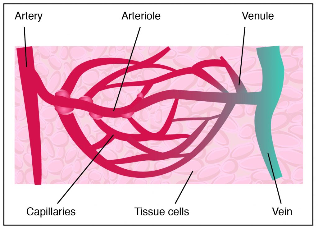
Because blood circulation is a closed loop system, blood is contained within the heart or blood vessels at all times. There are three types of blood vessels: arteries, veins, and capillaries. Arteries carry blood away from the heart, toward organs and tissues. Veins carry blood toward the heart, away from organs and tissues. Arteries branch into smaller blood vessels called arterioles, which further divide into capillaries. As shown in the following image, capillaries are tiny vessels that form a network around tissues. Veins branch into venules before further dividing into capillaries.

The heart is found between the lungs in the middle of the chest. It rests behind and slightly to the left of the sternum, or breastbone. The human heart is a muscular organ composed primarily of cardiac muscle. It consists of four chambers: two upper chambers called the atria and two lower chambers called the ventricles. The atria are separated from the ventricles by a muscular structure called the septum. Three layers make up the heart wall. These are the pericardium or outer layer, the myocardium or middle layer, and the endocardium or innermost layer. Most cardiac muscle tissue is found in the myocardium.

In addition to the four chambers, the heart has four valves that regulate blood flow into and out of the heart:
- Tricuspid valve regulates blood flow between the right atrium and right ventricle.
- Pulmonary valve regulates blood flow from the right ventricle into the pulmonary artery.
- Mitral valve regulates blood flow from the left atrium into the left ventricle.
- Aortic valve regulates blood flow from the left ventricle to the aorta. The aorta is the largest artery in the body.
Try this ATI TEAS science practice test question
Did You Know?

Capillaries have thin walls and a very large surface area. Because of the capillaries’ thin walls, blood flow slows to facilitate exchanges between blood and the body’s tissues.
ATI TEAS Science – Circulation and the Cardiac Cycle
Blood continually flows in one direction, beginning in the heart and proceeding to the arteries, arterioles, and capillaries. When blood reaches the capillaries, exchanges occur between blood and tissues. After this exchange happens, blood is collected into venules, which feed into veins and eventually flow back to the heart’s atrium. The heart must relax between two heartbeats for blood circulation to begin. Two types of circulatory processes occur in the body:

Systemic circulation
- The pulmonary vein pushes oxygenated blood into the left atrium.
- As the atrium relaxes, oxygenated blood drains into the left ventricle through the mitral valve.
- The left ventricle pumps oxygenated blood to the aorta.
- Blood travels through the arteries and arterioles before reaching the capillaries that surround the tissues.
Pulmonary Circulation

- Two major veins, the Superior Vena Cava and the Inferior Vena Cava, brings deoxygenated blood from the upper and lower half of the body.
- Deoxygenated blood is pooled into the right atrium and then sent into the right ventricle through the tricuspid valve, which prevents blood from flowing backward.
- The right ventricle contracts, causing the blood to be pushed through the pulmonary valve into the pulmonary artery.
- Deoxygenated blood becomes oxygenated in the lungs.
- Oxygenated blood returns from the lungs to the left atrium through the pulmonary veins.
Keep In Mind
Blood flow is regulated by many mechanisms in the body. This regulated variable is also directly proportional to blood pressure. If blood volume increases, blood pressure increases. The opposite occurs if blood pressure decreases.
The following image shows the heart’s role in systemic and pulmonary circulation.

The complete cycle beginning with atrial contraction and ending with ventricular contraction is called the cardiac cycle. When the heart contracts and pumps blood into systemic circulation, this is called systole. Diastole refers to the period of relaxation when the heart chambers fill with blood.
Because the heart is a muscle, it transmits electrical impulses that cause the heart to contract. This electrical activity can be recorded using an electrocardiogram, or EKG. An EKG is a graph that shows the heart’s rate and rhythm over a period of time. As shown in the following image of an EKG, waves in the graph have different meanings.

The first wave on an EKG is the P wave. This indicates atrial contraction or systole. The QRS complex represents the combination of Q, R, and S waves. This indicates ventricular systole or contraction. The T wave indicates ventricular diastole. The flat line between the S and T wave is the ST segment.
Let’s Review
- Blood is a type of connective tissue composed of formed elements, plasma, and other substances.
- Erythrocytes, leukocytes, and thrombocytes are the formed elements that make up blood.
- Blood transports substances throughout the body, regulates physiological processes, and protects the body.
- There are four common blood groups that are determined by inherited differences in antigens on red blood cells.
- Agglutination, or clumping, can be used to help interpret the blood type of a blood sample.
- The cardiovascular system circulates blood throughout the body in a closed loop structure.
- The heart is a muscular organ with four chambers: two atria and two ventricles.
- Deoxygenated blood flows through pulmonary circulation, and oxygenated blood flows through systemic circulation.
- The cardiac cycle refers to the contraction and relaxation states of the atria and ventricles.
- An electrocardiogram, or EKG, is used to record heart beat and rhythm.
The next ATI TEAS science lesson covers the respiratory system.
For Further Review and Studying
Make sure to answer a few cardiovascular system practice question bank questions to test your understanding of everything you have reviewed in this lesson. Each practice question has a video explanation from Nurse Gaelen to explain the question and concepts further. Answer at least six practice questions each time you review this lesson.
Cardiovascular System Flashcards
You May Subscribe to the online course to gain access to the full lesson content.
If your not ready for a subscription yet, be sure to check out our free practice tests and sample lesson at this link

